Stress echocardiography is a test that uses ultrasound imaging to show how well your heart muscle is working to pump blood to your body. It is most often used to detect a decrease in blood flow to the heart from narrowed coronary arteries. The test is well validated and used all over the world. In the UK there has been a large expansion in use over the last 10 years.
A new Stress echocardiography service has recently started at New Victoria Hospital with state of the art equipment
How the Test is Performed
This test is done in our Cardiac Room which is set up with appropriate equipment. The test is undertaken by a Consultant Cardiologist with support from a Cardiac Physiologist.
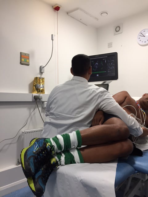
A resting echocardiogram will be done first. While you lie on your left side with your left arm out, a small ultrasound device called a transducer is held against your chest. A special gel is used to help the ultrasound waves get to your heart. After this you will have a 12 lead ECG which remains attached to your chest for the whole test.
Most people will walk on a treadmill (or pedal on an exercise bicycle) slowly and at 3 minute intervals you will be asked to walk faster and on a progressively steeper incline. In most cases, you will need to walk for around 6 to 15 minutes, depending on your level of fitness and your age. Your doctor will ask you to stop:
- When your heart is beating at the target rate
- When you are too tired to continue
- If you are having chest pain or a change in your blood pressure that worries the provider administering the test
- If the ECG demonstrates an abnormality
If you are not able to exercise, you will receive a drug called dobutamine via a cannula through a vein (intravenous line). This medicine will make your heart beat faster and harder, similar to when you exercise.
Your blood pressure and heart rhythm (ECG) will be monitored throughout the procedure.
More echocardiogram images will be taken while your heart rate is increasing, or when it reaches its peak. The images will show whether any parts of the heart muscle do not work as well when your heart rate increases. This is a sign that part of the heart may not be getting enough blood or oxygen because of narrowed or blocked arteries. The test can also identify any scarring from an old heart attack as well as assess the function of your heart valves.
Why the Test is Performed
The test is performed to see whether your heart muscle is getting enough blood flow and oxygen when it is working hard (under stress).
Your cardiologist may order a cardiac screening if you:
- Have symptoms of chest pain or shortness of breath
- Have recently had a heart attack
- Are going to have an operation and the surgeon or anaesthetist wants to be sure there are no issues with your heart
- You have had a coronary angiogram or CT coronary angiogram and the cardiologist wants to be sure a coronary narrowing identified is significant
- Have heart valve problems


